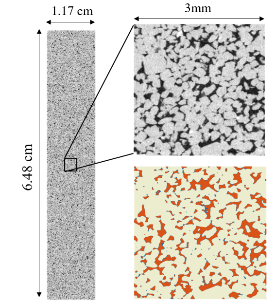
This dataset contains raw and segmented X-Ray micro-tomography images from steady-state drainage and imbibition flow experiments in two distinct Bentheimer sandstones. The dataset is presented and utilised in the paper 'Representative elementary volumes, hysteresis and heterogeneity in multiphase flow from the pore to continuum scale' available at: https://eartharxiv.org/2aejr/. Full details of the experimental setup, procedure and imaging analysis can be found in the paper and supporting information. The experiments consist of steady-state co-injection core floods with micro-CT imaging and simulatenous pressure measurments, performed on two Bentheimer sandstone cores. These cores have a diameter of 12.35mm (cored with a 12.7mm,1/2"drill bit), and length 73.2mm and 64.7mm, with core-averged porosities of 0.203 and 0.223 respectively. The shallow-marine Bentheimer sandstone typically consists of around 95% quartz with minor feldspar and clay and has a well-sorted grain size distribution. The first core is relatively homogeneous, whilst the second has oblique, low porosity layers orientated approximately 60 degrees to the flow direction. Brine (3.5wt.%KI) and decane (99.9%pure, Sigma-Aldrich) are used as wetting and non-wetting phases respectively, with an interfacial tension of appromxiately 51.16 mN/m (for the pure DI water - decanesystem). The outcrop Bentheimer is strongly water-wetting. The experiments are performed with a confining pressure of 4.5MPa, and pore-pressure of 1.5MPa. Scans are performed with a Zeiss Versa 510 X-Ray microscope with a flat panel detector and various microscope objectives. Firstly, a full-core dry scan is obtained, at a resolution of 6 microns. The core is then saturated with high salinity brine (30wt.%KI), and a full core scan obtained for differential imaging purposes. The working fluid brine (3.5wt.%KI) is then pumped through the core, fully displacing the high-salinity brine, and the absolute permeability is measured. Following this, decane and brine are co-injected into the core at constant total flow rate (0.1 ml/min) varying the fractional flow in a drainage and imbibition process. At each fractional flow, once the differential pressure has stabilised a full-core scan is taken followed by zoom-in, region of interest scans. For the 'full core' scans, 11 and 10 individual scans are taken at increasing vertical locations in each core, to cover a total volume after processing of 1950 x 1950 x 10800 voxels (11.7mm x 11.7mm x 64.8mm) and 1900 x 1900 x 9550 voxels (11.4mm x 11.4mm x 57.3mm) in core 1 and 2 respectively. Approximately 5mm is lost from the images near the inlet and outlet of the core due to noise from the end-pieces and the cone angle of the X-Ray beam. There is significant overlap between each scan to allow registration and merging of the full image. Each individual scan image is normalised to pre-selected references to obtain consistent grey-scale values throughout the core, registered with the first scan, and then merged to create a single image of the core. These are the 'raw' scans available in this data set. After each full-core scan, a selection of zoom-in, region of interest scans are performed to facilitate the interfacial curvature measurements in the paper. A 4x microscope objective lens is used to aquire each image, with the detector position varied to achieve a 3.5 micron resolution and 2 micron resolution image, each containing 1000 cubed voxels. These are the raw, ROI scans available in the dataset. The segmented images are also available in the data set for the full core and region of interest scans. The segmented images show the connected and disconnected oil for the full core scans (binary images with 1 being the oil), and grain, water and total oil for the region of the interest images. Alongside this, the segmented porosity map is also available, which was generated through segmentation of the differential image (high salinity and dry raw images). Details of the differential segmentation and watershed segmented used for the fluid phases can be found in the paper. The full core images herein have been divided into 50 equal subvolumes to faciliate downloading and utilising the dataset with small CPU and RAM requirements. The full, merged images can be found on the UKCCSRC data repository. All resolution data is contained within the file name: full_core means 6um, 3_45 means 3.45 microns, 3_5 means 3.5 microns and 2_00 means 2 microns. Absolute voxel sizes in the images may differ but can be changed in settings to match the given scan resolutions mentioned previously. Also contained in the file names are the oil fractional flow and flood process. DraFo05 refers to a drainage step with a fractional flow of oil of 0.05. ImbFo5 refers to a imbibition step with a fractional flow of oil of 0.5.
Samuel Jackson (Imperial College London)
Aug. 28, 2019
ODC-BY 1.0
10.17612/KT0B-SZ28
The downloadable archive contains all project data; the size of the archive file for this project is 728.12 GB.
Download